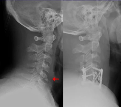
Open fracture
Open fractures involve a broken bone protruding through the skin.

Alan Hughes · CC BY-SA 2.0
An open fracture, also called a compound fracture, is a type of bone fracture where the broken bone creates an open wound in the skin, often by piercing through the surface. This injury can be life-threatening or limb-threatening due to risks of deep infection and bleeding, and is frequently caused by high-energy trauma such as road traffic accidents.
- most_commonly_injured_bone
- tibia
- highest_risk_group
- working-age young men
- male_to_female_ratio
- 7 to 3
- mean_age_in_males
- 40.8 years
- mean_age_in_females
- 56 years
- infection_rate_grade_I
- 1.4%
Lore & Background
Open fractures are often caused by high-energy trauma such as road traffic accidents, firearms, and falls from height. The severity varies widely, from a small 'poke-hole' wound to extensive soft tissue damage involving nerves, tendons, muscles, and blood vessels. The Gustilo-Anderson open fracture classification is the most commonly used system for diagnosis, treatment guidance, and outcome prediction, though it has limited interobserver reliability (50% to 60%) and can only be accurately graded in the operating theatre.
Reader's Guide
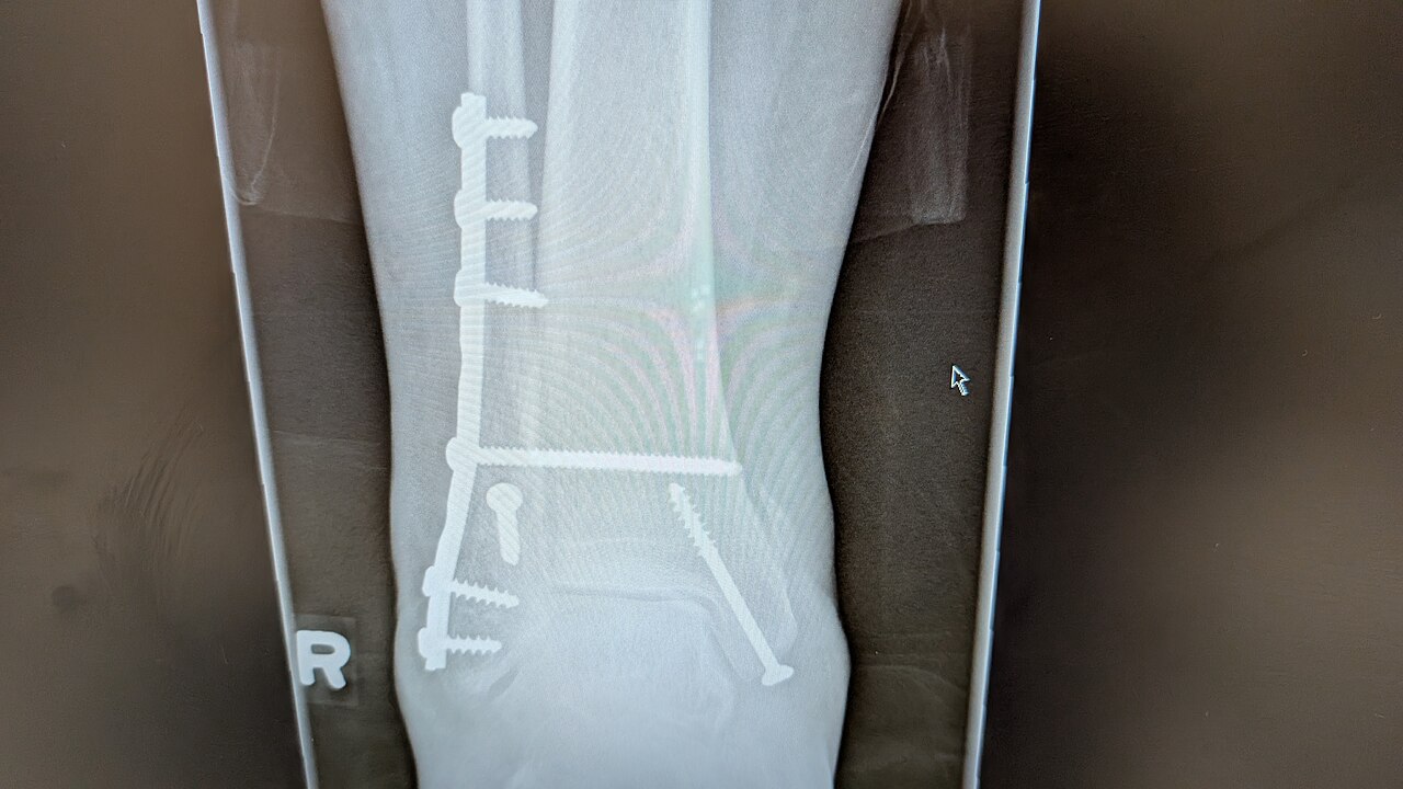
The management of open fractures begins with Advanced Trauma Life Support to rule out other life-threatening injuries. Antibiotics, typically first-generation cephalosporins sometimes combined with aminoglycosides, are administered as soon as possible, ideally within an hour, and continued for at least three days for higher-grade fractures. Core principles include therapeutic irrigation, wound debridement, early wound closure, and bone fixation to reduce infection risk and promote healing. Tetanus prophylaxis is routinely given, with anti-tetanus immunoglobulin reserved for highly contaminated wounds. The decision to attempt limb salvage rather than amputation should involve a full discussion between doctors, the patient, and their family, not solely based on injury severity scores.
Did You Know?
- Road traffic accidents are the most common cause of open fractures, followed by falls from height and crush injuries.
- Infection rates range from 1.4% for Gustilo Grade I fractures to 10–50% for Grade IIIB and IIIC fractures.
- Pre-operative wound cultures are no longer recommended because they are positive in only 22% of pre-debridement samples.
- Antibiotic-impregnated devices such as tobramycin-impregnated PMMA beads can help reduce infection risk.
The Mechanism Behind the Dinner Fork
A Colles' fracture is most often the result of a person falling onto a hard surface and instinctively bracing with an outstretched hand. The force drives the distal radius backward, and if the wrist is flexed at impact, the injury shifts toward what is known as a Smith's fracture instead. The break typically lands three to five centimetres above the radio-carpal joint, with the broken fragment displacing posteriorly and laterally. This produces the unmistakable silhouette that has given the injury its colorful nicknames: the "dinner fork" or "bayonet" deformity. Patients present with pain, swelling, visible deformity, and bruising around the wrist. In some cases the tip of the ulna fractures as well, and the median nerve can sustain damage as a complication. The fracture is the second most common osteoporosis-related break, trailing only vertebral fractures, which underscores how bone density plays a central role in who is vulnerable to this particular injury.
Reading the Fracture on Film
Confirming a Colles' fracture generally requires only anteroposterior and lateral X-ray views, though the classic presentation includes a transverse break roughly two and a half centimetres proximal to the radio-carpal joint, accompanied by dorsal displacement, dorsal angulation, and radial tilt. Additional hallmarks that radiologists look for include radial shortening, loss of ulnar inclination, radial angulation of the wrist, and comminution at the fracture site. Notably, more than sixty percent of cases also involve a fracture of the ulnar styloid process. The term "Colles fracture" was originally reserved for a break at the cortico-cancellous junction of the distal radius, but modern usage has broadened it to encompass any distal radius fracture with dorsal displacement, regardless of whether the ulna is involved. Several formal classification schemes exist to grade severity and guide treatment, including the Frykman, Gartland and Werley, Lidström, Nissen-Lie, and Older's systems. Colles himself, writing in 1814, described the injury as occurring about an inch and a half above the carpal extremity of the radius, with the carpus thrown backward.
From Cast to Operating Table
Treatment for a Colles' fracture is tailored to how displaced and complex the break is. A stable, undisplaced fracture can be managed with a simple cast, applied with the distal fragment held in palmar flexion and ulnar deviation. Mildly angulated fractures may call for a closed reduction, during which procedural sedation, analgesia, or a hematoma block keeps the patient comfortable. For significant angulation or deformity, surgeons resort to open reduction with internal fixation or external fixation. A volar forearm splint serves as the go-to temporary immobilization device while the definitive plan is arranged. Several instability criteria help determine whether surgery is warranted: dorsal tilt exceeding twenty degrees, comminution, abruption of the ulnar styloid, intra-articular displacement greater than one millimetre, or loss of radial height beyond two millimetres. The more of these criteria present, the stronger the case for operative intervention. In patients over fifty, surgical reduction followed by casting is feasible in the majority of cases. Follow-up X-rays at one, two, and six weeks verify that healing is progressing correctly, and full recovery can stretch from two months to a year or longer.
An Irish Surgeon's Legacy and the People at Risk
The fracture bears the name of Abraham Colles, an Irish surgeon from Kilkenny who lived from 1773 to 1843. In 1814, long before X-rays existed, he identified and described the injury purely by observing the characteristic backward deformity of the forearm. Ernest Amory Codman later pioneered the use of radiography to study the fracture, publishing his findings in the Boston Medical and Surgical Journal, the predecessor of The New England Journal of Medicine, and developing an early classification system. Though Claude Pouteau is sometimes credited as the first describer, the orthopaedic historian P. Liverneaux disputes that claim. Epidemiologically, roughly fifteen percent of people will sustain a Colles' fracture at some point in their lives. Women are disproportionately affected, largely because of post-menopausal osteoporosis. The injury clusters in young adults and the elderly, while children and middle-aged adults are less commonly involved. In older patients the weaker cortex tends to produce extra-articular breaks, whereas younger individuals, requiring greater force to fracture, often sustain more complex intra-articular patterns. In children with open growth plates, the equivalent injury manifests as an epiphyseal slip, classified as a Salter type I or II fracture.
Gallery
Frequently Asked Questions
What is Open fracture?
An open fracture, also known as a compound fracture, occurs when a broken bone pierces through the overlying skin, creating a wound that exposes the bone to the outside environment. It is most often the result of high-energy trauma, such as a severe road traffic accident.
What are Open fracture's main threats?
The two greatest dangers are deep bone infection and uncontrolled bleeding, which together can make the injury either limb-threatening or life-threatening. The tibia is the bone most frequently involved in this type of injury.
Who is most likely to encounter Open fracture?
Working-age young men make up the highest-risk demographic, with a male-to-female ratio of roughly 7 to 3. The average age at injury sits around 40.8 years for men and 56 years for women.
How does Open fracture's story end?
With urgent surgical debridement, stabilization, and appropriate antibiotics, most patients go on to heal, though the risk of infection depends on the injury's grade. For the mildest Grade I open fractures, the infection rate is only about 1.4%.
Why is Open fracture important in orthopedics?
It is treated as a surgical emergency because the exposed bone and soft tissues are immediately vulnerable to contamination and rapid, deep-seated infection. Unlike a closed fracture, it demands swift intervention to preserve the limb and prevent systemic complications.
More in Musculoskeletal System And Orthopedics 1-17
Spotted an error? Know more?
This is a living reference — every entry is fact-audited, and reader corrections feed straight into our audit queue. Suggest an edit · See this site's audit record