Shoulder
The shoulder is the most mobile and potentially unstable joint.

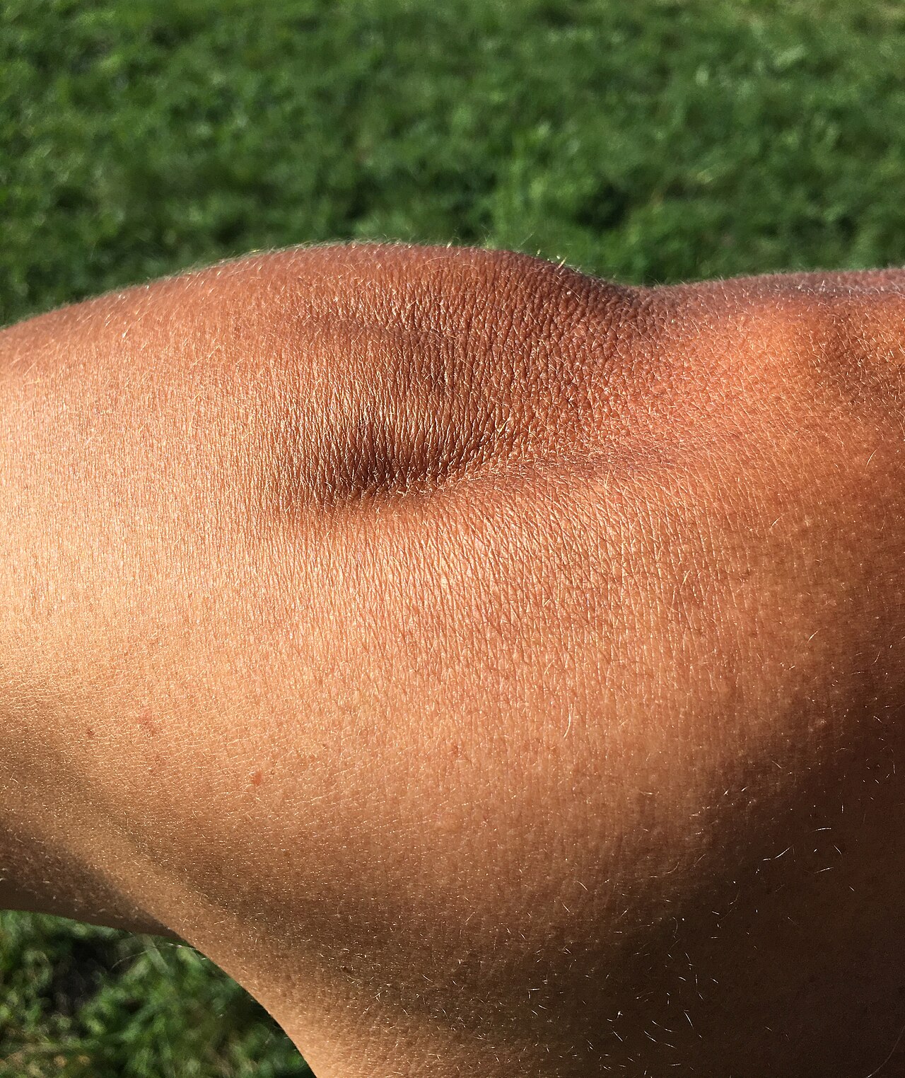
Elements Of This World · CC BY 2.0
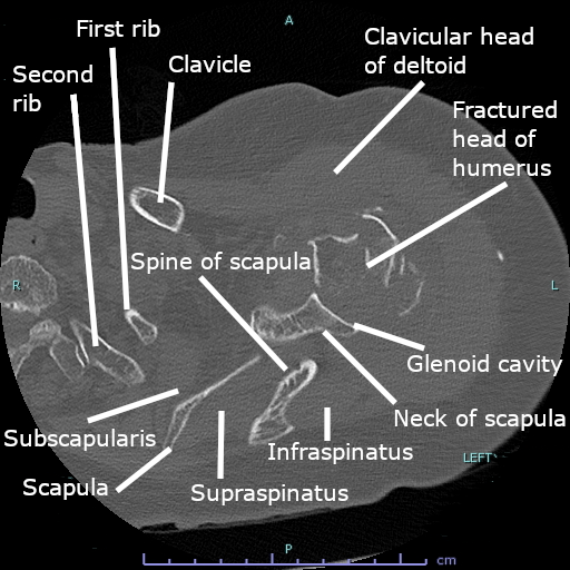
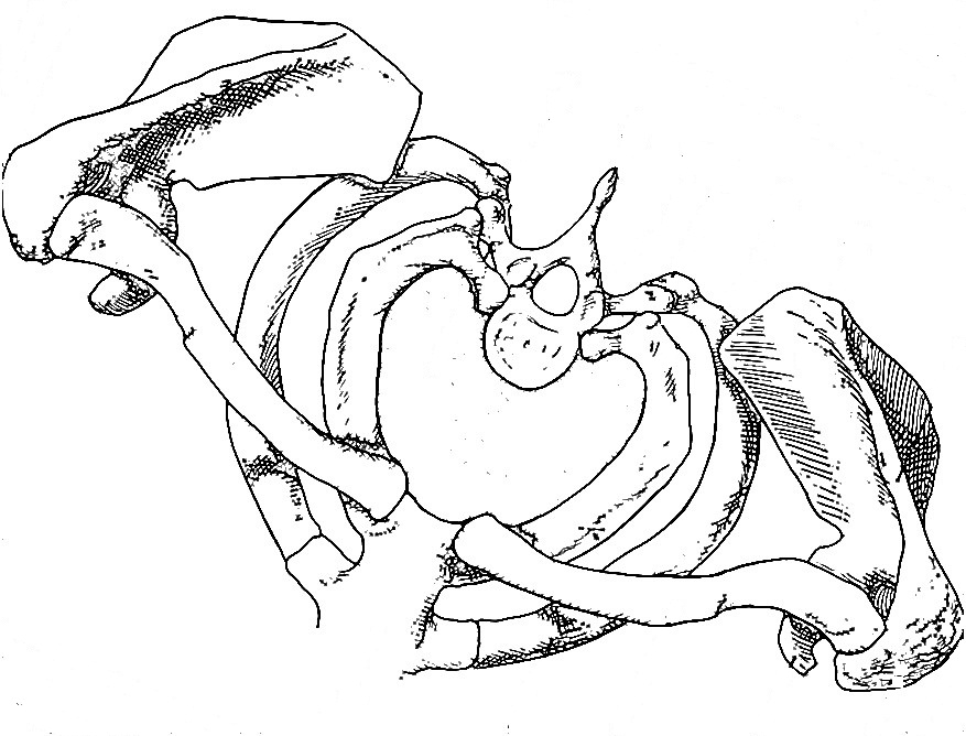
The shoulder is the group of structures in the region of the joint where the humerus attaches to the scapula. It is made up of three bones: the clavicle, the scapula, and the humerus, as well as associated muscles, ligaments, and tendons. The shoulder joint, also known as the glenohumeral joint, is the major joint of the shoulder and is a ball-and-socket joint that allows the arm to rotate in a circular fashion or hinge out and up away from the body.
- bones
- clavicle, scapula, humerus
- major_joint
- glenohumeral joint
- joint_type
- ball and socket
- rotator_cuff_muscles
- supraspinatus, subscapularis, infraspinatus, teres minor
- stability
- extremely unstable, prone to dislocation
Lore & Background
The shoulder consists of a ball-and-socket joint formed by the humerus and scapula, with the head of the humerus sitting in the glenoid cavity. The glenoid cavity is shallow, with a 4-to-1 disproportion in size between the large humeral head and the cavity, which is deepened by the fibrocartilaginous glenoid labrum. The joint capsule is a soft tissue envelope lined by a synovial membrane, strengthened by ligaments including the coracohumeral and glenohumeral ligaments.
Reader's Guide
The shoulder's significance lies in its balance between mobility and stability. It must be mobile enough for the wide range of actions of the arms and hands, yet stable enough for lifting, pushing, and pulling. The rotator cuff, a group of four muscles, directly surrounds and attaches to the shoulder joint, holding the humeral head in the glenoid cavity during movement. The armpit (axilla) is formed by the space between shoulder muscles and contains nerves, blood vessels, and lymph nodes. The brachial plexus supplies most shoulder muscles, and blood is supplied by the axillary artery and branches of the thyrocervical trunk.
Did You Know?
- The shoulder joint has an approximately 4-to-1 disproportion in size between the large head of the humerus and the shallow glenoid cavity.
- The rotator cuff consists of four muscles: supraspinatus, subscapularis, infraspinatus, and teres minor.
- Two filmy sac-like structures called bursae permit smooth gliding between bone, muscle, and tendon in the shoulder.
- The glenoid labrum is a more fibrous or rigid cartilage found only around the socket of the shoulder joint.
Anatomy & Biomechanics: Freedom at a Cost
The shoulder stands as the body's most mobile joint, a distinction that comes at a significant cost. Built from three bones—the collarbone, shoulder blade, and upper arm bone—it operates through two primary articulations. The acromioclavicular joint connects the highest point of the shoulder blade to the collarbone, while the glenohumeral joint, the one most people mean when they say "shoulder," functions as a ball-and-socket mechanism. The rounded head of the humerus nests into a shallow dish on the scapula's outer edge, permitting circular rotation and wide lateral movement. The scapula's own ability to glide along the rib cage adds yet another layer of mobility. This extraordinary freedom, however, renders the joint inherently unstable. A soft tissue capsule lined with a smooth synovial membrane wraps the glenohumeral articulation, while three ligaments anchor its front. Tendons connect muscle to bone for movement, and four small bursa sacs provide cushioning between the rotator cuff and the bony arch above it. The trade-off is clear: maximum range of motion demands a structure vulnerable to wear, breakdown, and injury.
The Rotator Cuff: Four Muscles, Four Vulnerabilities
The rotator cuff, a composite structure of four tendons and their associated muscles, serves as the shoulder's primary stabilizer, holding the rounded head of the humerus securely within its shallow glenoid socket while simultaneously enabling powerful, controlled movement. The four members—supraspinatus, infraspinatus, teres minor, and subscapularis—can be remembered with the handy acronym S.I.T.S. Each muscle bears a distinct mechanical role and, consequently, a distinct vulnerability to injury. The supraspinatus, the most frequently damaged of the four, assists in lifting the arm away from the body; it is susceptible to repetitive overhead work, subacromial impingement, or a fall on an outstretched hand. The infraspinatus and teres minor both externally rotate the humerus. The former is commonly strained during the deceleration phase of a throwing motion, while the latter is stressed by excessive external rotation, a pattern common among overhead athletes. The subscapularis, an internal rotator, can be over-stretched by forced external rotation. Injuries to the cuff span a wide severity spectrum, ranging from mild tendonitis and joint instability all the way to complete tears that demand surgical intervention.
Diagnosis: Listening, Touching, and Ruling Out the Rest
Diagnosing shoulder problems begins with a careful history and physical examination. Clinicians gather details on the patient's age, dominant hand, whether the issue disrupts daily work, and whether symptoms are acute or chronic. They probe for catching, locking, instability, burning sensations, stiffness, swelling, and weakness. A structured framework called OPQRST—covering onset, provocation, quality, radiation, severity, and timing—helps organize the patient's narrative. Crucially, doctors must consider that pain felt at the shoulder may actually originate elsewhere: the gallbladder, liver, heart, cervical spine, peptic ulcer, or even pneumonia can all produce sensations the brain interprets as shoulder pain. The physical exam proceeds through inspection for asymmetry, atrophy, bruising, or swelling; palpation across the glenohumeral joint, acromioclavicular joint, biceps tendon, and surrounding structures; and range-of-motion testing in rotation, abduction, and adduction. The Apley scratch test, in which the patient reaches behind the head and behind the back, is considered the most useful single maneuver. Standardized tools like the Penn Shoulder Score further quantify pain and function for tracking over time.
Recovery Beyond the Tear: Rehabilitation and New Frontiers
Recovering shoulder function is not simply a matter of waiting for damaged tissue to heal on its own. Evidence indicates that restoring full range of motion, rebuilding muscular strength, and retraining coordinated motor control through structured rehabilitation are central to a patient's return to meaningful function. This insight shifts the clinical focus from purely structural repair to active, guided movement and neuromuscular re-education. Recent advances have introduced virtual reality–based therapy as a promising complementary approach to traditional physical therapy. By immersing patients in interactive, game-like exercises, VR systems have been shown to boost engagement and motivation during the repetitive shoulder movements that form the backbone of rehabilitation. Studies indicate these programs can improve specific outcomes such as abduction range of motion while achieving pain relief and overall functional gains comparable to conventional therapy. The appeal lies in making the often tedious process of repeated range-of-motion work feel purposeful and engaging, potentially accelerating the very restoration of mobility and strength that underpins a successful shoulder recovery.
Gallery
Frequently Asked Questions
What is the Shoulder in the musculoskeletal canon?
The Shoulder is the anatomical region where the humerus meets the scapula, formed by three bones—the clavicle, scapula, and humerus—along with their supporting muscles, ligaments, and tendons. It serves as the primary connection point that lets the upper limb move freely relative to the torso.
What bones make up the Shoulder?
Exactly three bones form the Shoulder: the clavicle (collarbone), the scapula (shoulder blade), and the humerus (upper-arm bone). These are linked by muscles, ligaments, and tendons that coordinate movement and provide structural support.
What is the Shoulder's major joint and how does it move?
The glenohumeral joint is the Shoulder's principal articulation, classified as a ball-and-socket joint. This design permits the arm to rotate in a full circle as well as hinge outward and upward away from the body.
Which muscles act as the Shoulder's rotator cuff?
Four muscles form the rotator cuff: the supraspinatus, subscapularis, infraspinatus, and teres minor. Together they stabilize the humeral head within the shallow glenoid cavity while the arm moves.
Why is the Shoulder considered the most unstable joint in the body?
Because the glenohumeral socket is very shallow relative to the humeral head, the joint sacrifices bony constraint for maximum range of motion. This makes it the most mobile yet most dislocation-prone joint in the human skeleton.
More in Musculoskeletal System And Orthopedics 1-17
Spotted an error? Know more?
This is a living reference — every entry is fact-audited, and reader corrections feed straight into our audit queue. Suggest an edit · See this site's audit record