Sprain
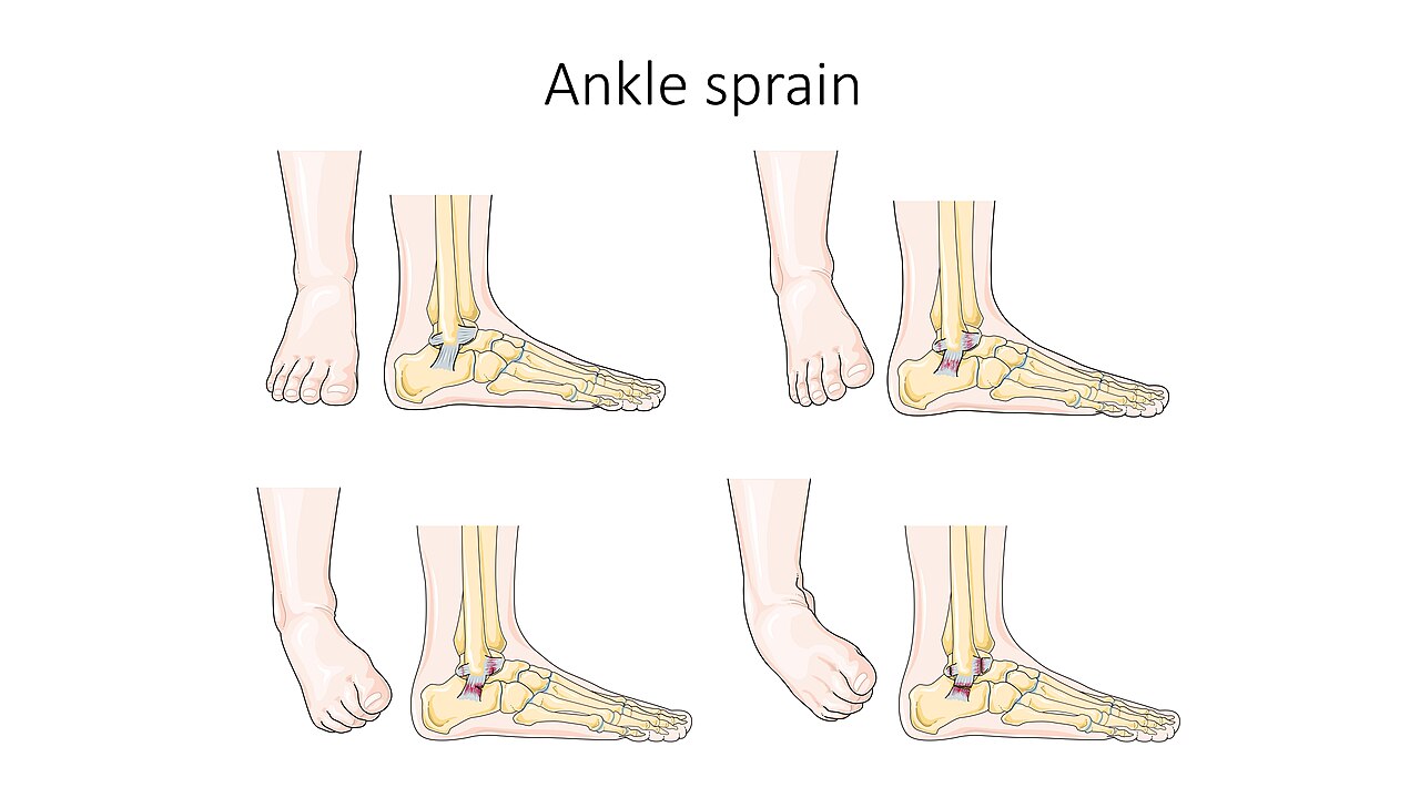
Ligament injury from joint overextension; common in ankles, knees, wrists.

Chinissai · CC BY-SA 3.0
A sprain is a soft tissue injury of the ligaments within a joint, often caused by a sudden movement abruptly forcing the joint to exceed its functional range of motion. Ligaments are tough, inelastic fibers made of collagen that connect two or more bones to form a joint and are important for joint stability and proprioception, which is the body's sense of limb position and movement.
- classification
- First degree (mild), second degree (moderate), third degree (severe)
- common sites
- Ankle, knee, wrist
- primary cause
- Repetitive movements (overuse) or acute trauma
- typical treatment
- RICE (rest, ice, compression, elevation) for mild cases; surgery for severe
- equivalent injury
- Strain (to muscle or tendon)
Lore & Background
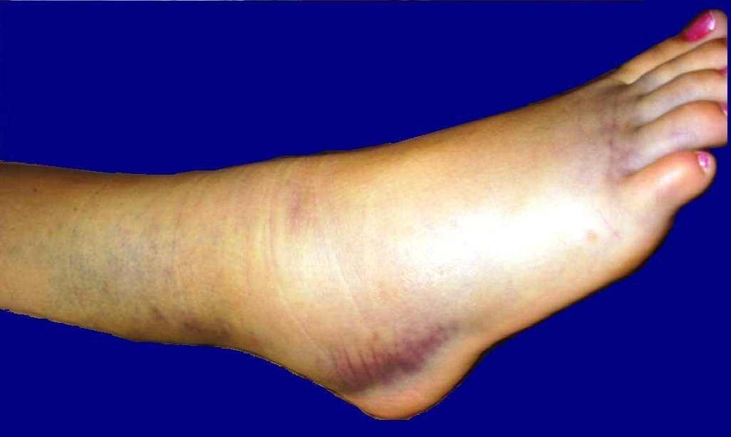
Sprains occur when a joint is forced beyond its functional range of motion, causing stretching or tearing of collagen fibers in ligaments. The majority of sprains are mild, with minor swelling and bruising that resolve with conservative treatment such as RICE. However, severe sprains involve complete tears or avulsion fractures, requiring surgical fixation and prolonged rehabilitation.
Reader's Guide
Sprains are among the most common musculoskeletal injuries, particularly affecting the ankle, knee, and wrist. Their significance lies in the role of ligaments in joint stability and proprioception; injury can lead to chronic instability if not properly managed. Diagnosis is often clinical, supplemented by X-ray or MRI when fractures or severe soft tissue damage are suspected. Treatment ranges from rest and ice to surgery for complete ruptures. Understanding the classification—first, second, or third degree—helps guide prognosis and recovery time. The distinction from strains (muscle or tendon injuries) and fractures is critical for appropriate care.
Did You Know?
- Ligaments have only about a 4% elastic zone before fibers rupture.
- Intra-articular ligaments have diminished blood flow compared to extra-capsular or capsular ligaments, affecting healing.
- Normal daily activity maintains about 80–90% of a ligament's mechanical properties.
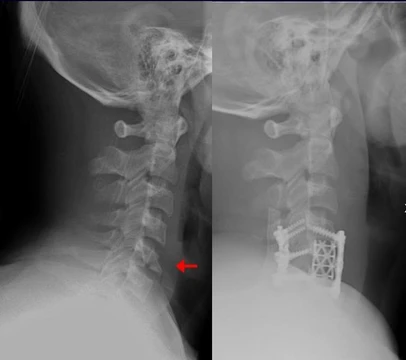
- A severe sprain may involve an avulsion fracture, where a piece of bone is pulled off.
The Mortise: A Unique Skeletal Lock
The ankle stands apart from every other joint in the human skeleton as the sole example of a mortise-and-tenon articulation, a name borrowed from woodworking that captures how the talus sits snugly within a rectangular socket formed by the tibia and fibula. The articular surface of the tibia is known as the plafond, a French word meaning ceiling, while the bony knobs extending downward from the inner tibia and the outer fibula are called the medial and lateral malleoli respectively. Together these structures cradle the talus and, with the help of supporting ligaments, keep it locked beneath the tibia. The tibia-talus contact carries the greater share of body weight compared to the smaller fibula-talus interface. Every bony surface in the ankle is lined with articular cartilage, and the gaps between the bones are remarkably tight: roughly 1.70 millimeters between the talus and medial malleolus, about 2.04 millimeters to the tibial plafond, and 2.13 millimeters to the lateral malleolus. Clinically, a measurable narrowing of these gaps signals the onset of osteoarthritis.
Three Joints, One Region
In everyday language the word ankle simply names the area where the leg meets the foot, but in medical terminology it can mean either that broad region or specifically the talocrural joint. The region itself stretches distally from the narrowest point of the lower leg and encompasses the portions of the foot nearest the body, up to the heel and the dorsal surface. Within this compact zone sit three distinct articulations: the talocrural joint proper, the subtalar or talocalcaneal joint, and the inferior tibiofibular joint. The talocrural joint is a synovial hinge responsible for the two primary movements of the foot—dorsiflexion and plantarflexion. When the foot is plantarflexed, the joint also permits a degree of side-to-side gliding, rotation, adduction, and abduction. Because the subtalar joint plays a major role in orienting the foot in space, some anatomists label it the lower ankle joint and reserve the term upper ankle joint for the talocrural articulation, a distinction that underscores how much functional complexity is packed into a space barely wider than a hand.
Ligaments and the Logic of the Sprain
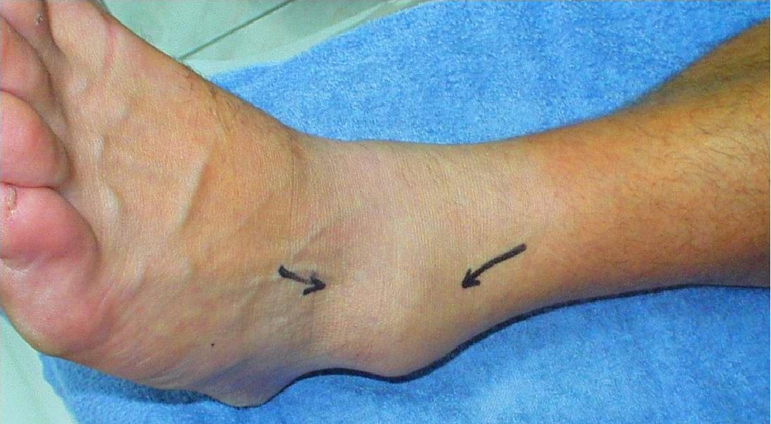
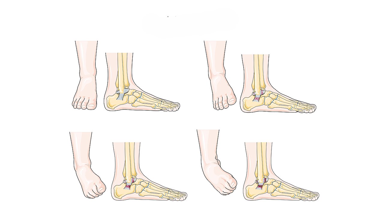
The ankle's stability depends on a carefully arranged set of ligaments. On the medial side, the robust deltoid ligament anchors at the medial malleolus and fans out to attach at four separate points on the calcaneus, navicular, and talus. The lateral side is guarded by three ligaments: the anterior talofibular, the posterior talofibular, and the calcaneofibular, all originating from the lateral malleolus. An additional player, the syndesmotic ligament, spans the articulation between the distal fibula and tibia; an isolated tear here is what clinicians call a high ankle sprain. The bony architecture of the mortise is inherently most stable when the foot is dorsiflexed, which means that in plantarflexion the ligaments must do considerably more of the work. This is precisely why the classic inversion sprain—where the anterior talofibular ligament is the most frequently damaged structure—tends to occur with the foot pointed downward. In severe cases the calcaneofibular ligament is also torn, compounding the instability.
The Tendon Corridor and Its Guardians
A dense bundle of tendons, arteries, veins, and nerves threads through the ankle region, and the body has built a series of connective-tissue bands called retinacula to keep them in place. Without these straps, the tendons would bowstring away from the bony angle between leg and foot, losing their mechanical advantage. On the front of the ankle, the superior extensor retinaculum spans the anterior surfaces of the tibia and fibula, housing the anterior tibial vessels, the tibialis anterior tendon, and the unsheathed tendons of extensor hallucis longus and extensor digitorum longus, while the deep and superficial peroneal nerves pass beneath and outside it respectively. Below, the Y-shaped inferior extensor retinaculum anchors to the calcaneus and blends upward with its superior counterpart. On the medial side, the flexor retinaculum runs from the medial malleolus to the medial process of the calcaneus, guiding the tibialis posterior, flexor digitorum longus, posterior tibial vessels, tibial nerve, and flexor hallucis longus in strict medial-to-lateral order. Laterally, the fibular retinacula secure the fibularis longus and brevis tendons against the outer ankle.
Gallery

Frequently Asked Questions
Who is Sprain?
Sprain is a soft-tissue injury in which the collagen fibers of a joint's ligaments are stretched or torn past their normal working range, usually triggered by a sudden, forceful motion. It most commonly strikes the ankle, knee, and wrist joints.
What are Sprain's powers/role?
Ligaments are the tough, non-stretchy collagen bands that anchor two or more bones together at a joint, acting as the body's mechanical stabilizers. They also relay positional feedback to the nervous system so the brain can track where each limb is in space.
How does Sprain's story end?
Mild (first-degree) sprains typically heal with the RICE protocol—rest, ice, compression, and elevation—over a few weeks. Third-degree (complete) tears, however, often require surgical repair followed by a structured rehabilitation program.
Why is Sprain important?
Ligament damage directly undermines joint stability and the proprioceptive signals that guide coordinated movement, making sprains a major concern in both sports medicine and everyday injury care. Left untreated, chronic instability can cascade into long-term joint dysfunction.
What's the difference between Sprain and Strain?
A sprain specifically involves ligament fibers at a joint, whereas a strain targets the muscle belly or its attached tendon. They are frequently confused in casual conversation, but they affect different tissue types even though both arise from overstretching.
More in Musculoskeletal System And Orthopedics 1-17
Spotted an error? Know more?
This is a living reference — every entry is fact-audited, and reader corrections feed straight into our audit queue. Suggest an edit · See this site's audit record